Home

Trompete Null Tofu allium cepa mikroskop Schöne Frau Schiffsform Verknüpfung

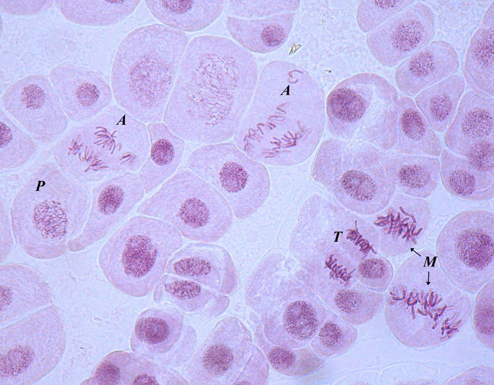
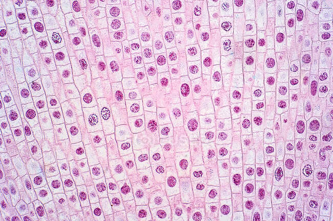
Lichtmikroskopie von Zwiebel- (Allium cepa) Wurzelspitzenzellen, die einer  Mitose (Kernteilung) unterzogen werden). — Schule, nuklear - Stock Photo |  #527495868
Lichtmikroskopie von Zwiebel- (Allium cepa) Wurzelspitzenzellen, die einer Mitose (Kernteilung) unterzogen werden). — Schule, nuklear - Stock Photo | #527495868

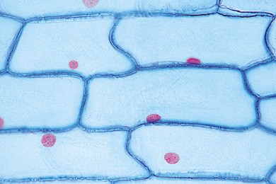
Zwiebel-epidermis Mit Großen Zellen Unter Dem Lichtmikroskop. Klare  Epidermale Zellen Einer Zwiebel, Allium Cepa, In Einer Einzigen Schicht.  Jede Zelle Mit Wand, Membran, Zytoplasma, Kern Und Großer Vakuole. Foto.  Lizenzfreie Fotos, Bilder
Zwiebel-epidermis Mit Großen Zellen Unter Dem Lichtmikroskop. Klare Epidermale Zellen Einer Zwiebel, Allium Cepa, In Einer Einzigen Schicht. Jede Zelle Mit Wand, Membran, Zytoplasma, Kern Und Großer Vakuole. Foto. Lizenzfreie Fotos, Bilder
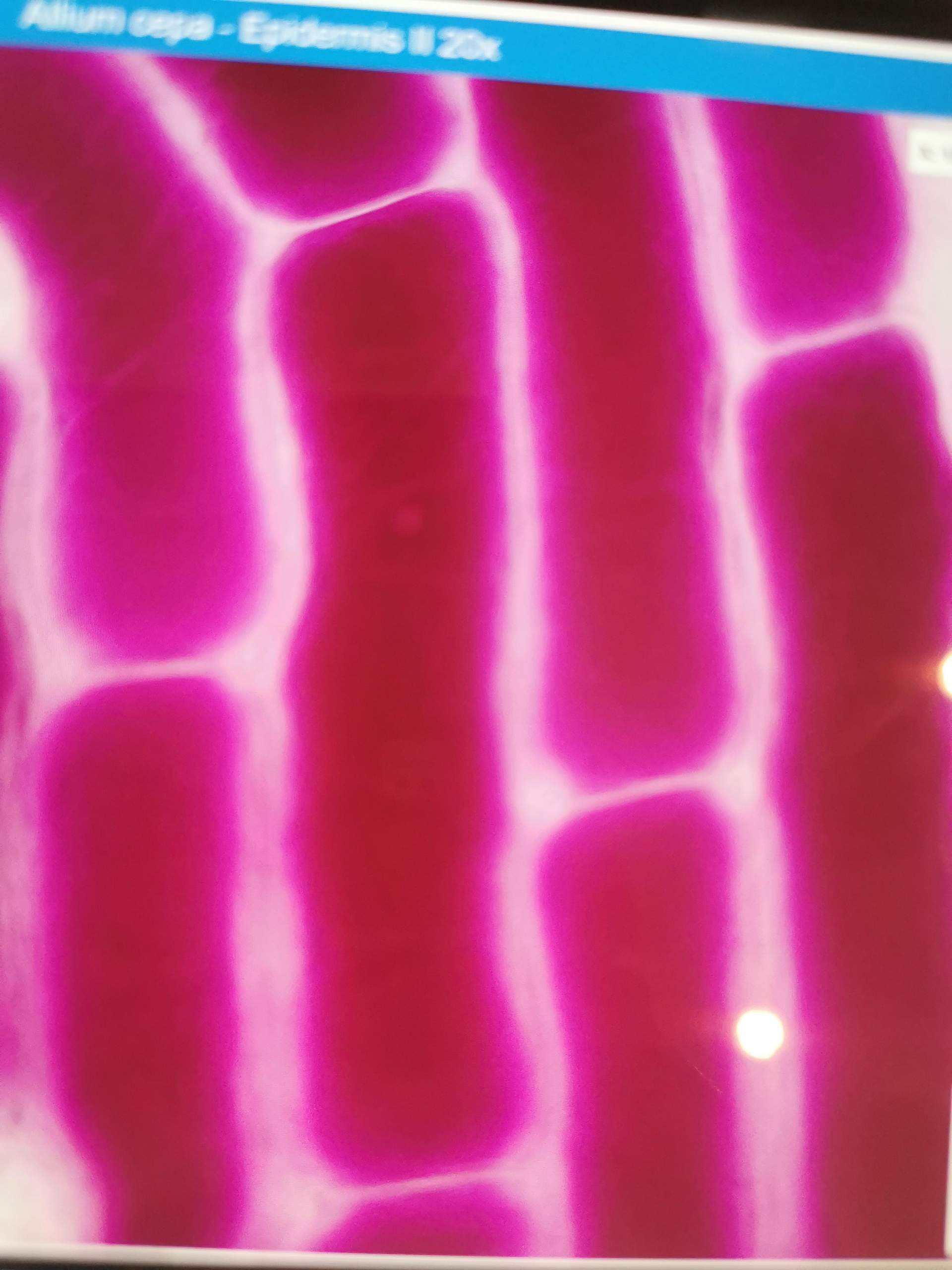
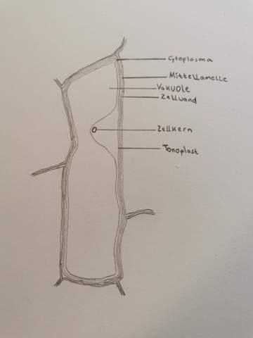
Botanische Mikroskopie Zeichnung wo Zeichne ich das Cytoplasma. Allium Cepa?  (Botanik, Zellen, zellkern)
Botanische Mikroskopie Zeichnung wo Zeichne ich das Cytoplasma. Allium Cepa? (Botanik, Zellen, zellkern)

Zwiebelepidermis (Allium cepa) mit Zellen und Zellkern. Optisches Mikroskop  X200 Stockfotografie - Alamy
Zwiebelepidermis (Allium cepa) mit Zellen und Zellkern. Optisches Mikroskop X200 Stockfotografie - Alamy
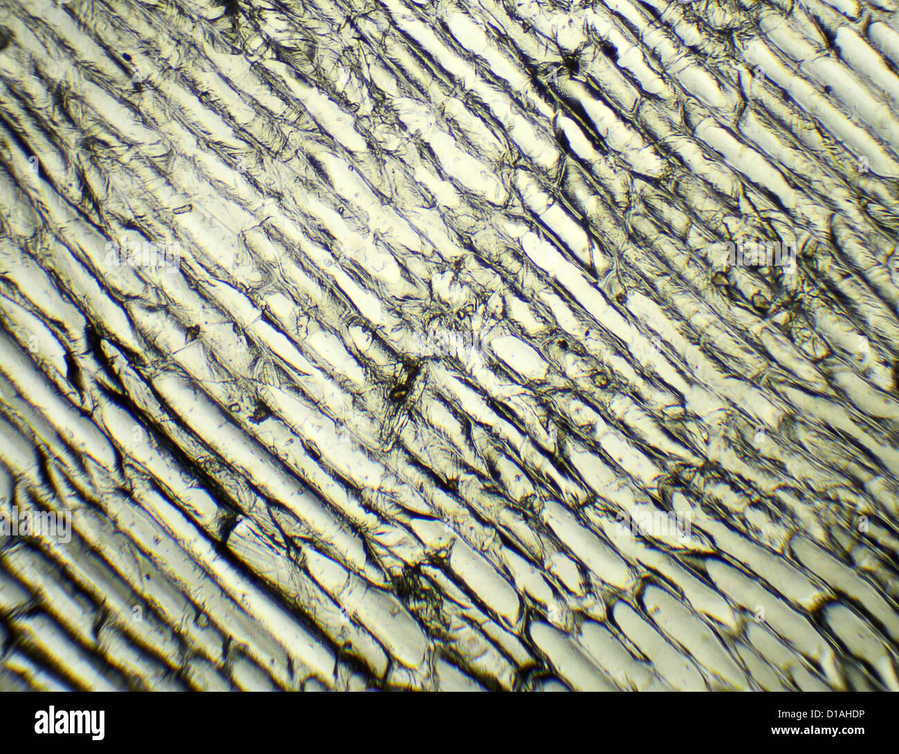
Zwiebel unter dem Mikroskop, Hintergrund. (Allium Cepa Stockfotografie -  Alamy
Zwiebel unter dem Mikroskop, Hintergrund. (Allium Cepa Stockfotografie - Alamy

Bawang Merahhh | PDF

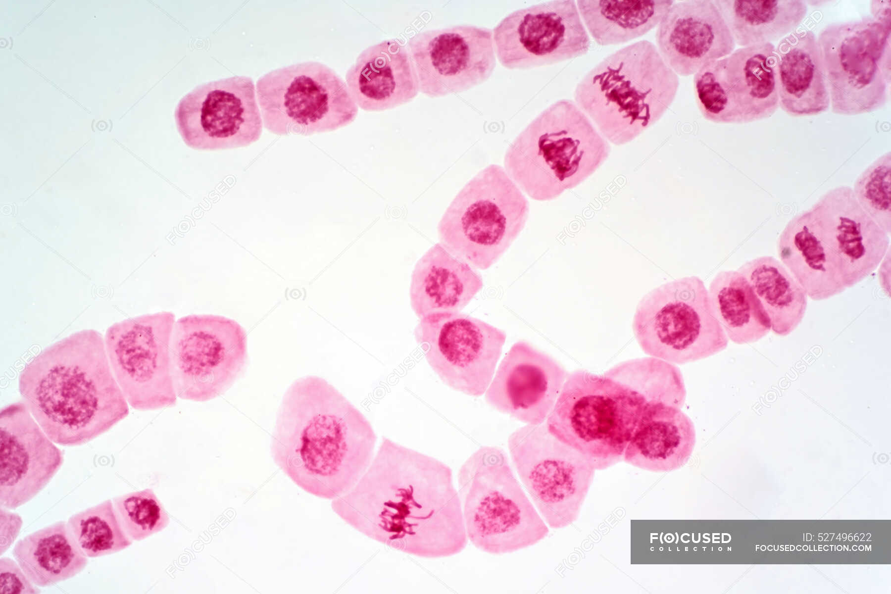
Lichtmikroskopie von Zwiebel- (Allium cepa) Wurzelspitzenzellen, die einer  Mitose (Kernteilung) unterzogen werden). — Abschnitt, nuklear - Stock Photo  | #527496622
Lichtmikroskopie von Zwiebel- (Allium cepa) Wurzelspitzenzellen, die einer Mitose (Kernteilung) unterzogen werden). — Abschnitt, nuklear - Stock Photo | #527496622
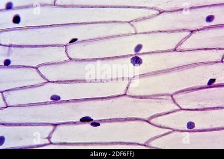
Allium cepa, Küchenzwiebel, Epidermis total. | LMS Lehrmittel-Service  H.Späth GmbH
Allium cepa, Küchenzwiebel, Epidermis total. | LMS Lehrmittel-Service H.Späth GmbH

Zwiebel unter dem Mikroskop, Hintergrund. (Allium Cepa Stockfotografie -  Alamy
Zwiebel unter dem Mikroskop, Hintergrund. (Allium Cepa Stockfotografie - Alamy

Zwiebelepidermis (Allium cepa) mit Zellen und Zellkern. Optisches Mikroskop  X100 Stockfotografie - Alamy
Zwiebelepidermis (Allium cepa) mit Zellen und Zellkern. Optisches Mikroskop X100 Stockfotografie - Alamy
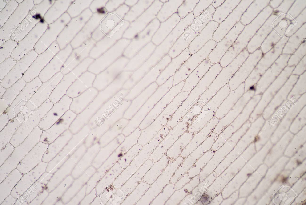
Cells Of The Onion Skin - Allium Cepa Microscope. Stock Photo, Picture And  Royalty Free Image. Image 44129708.
Cells Of The Onion Skin - Allium Cepa Microscope. Stock Photo, Picture And Royalty Free Image. Image 44129708.

Epidermiszellen Zwiebelschale - Karl Kaiser Website

Botanik online: Intrazelluläre Strukturen - Zellkern von Allium cepa - UV- Mikroskopie

Allium cepa root tip by Cursed-9-11 on DeviantArt

Zwiebel Epidermis Mit Großen Zellen Unter Dem Mikroskop Stockfoto und mehr  Bilder von Haut - iStock
Zwiebel Epidermis Mit Großen Zellen Unter Dem Mikroskop Stockfoto und mehr Bilder von Haut - iStock

Rote Zwiebeln Peeling Unter Dem Mikroskop Stockfoto und mehr Bilder von  Zelle - Zelle, Zwiebel, Schale - iStock
Rote Zwiebeln Peeling Unter Dem Mikroskop Stockfoto und mehr Bilder von Zelle - Zelle, Zwiebel, Schale - iStock

Zwiebel-epidermis Mit Großen Zellen Unter Dem Lichtmikroskop. Klare  Epidermale Zellen Einer Zwiebel, Allium Cepa, In Einer Einzigen Schicht.  Jede Zelle Mit Wand, Membran, Zytoplasma, Kern Und Großer Vakuole. Foto.  Lizenzfreie Fotos, Bilder
Zwiebel-epidermis Mit Großen Zellen Unter Dem Lichtmikroskop. Klare Epidermale Zellen Einer Zwiebel, Allium Cepa, In Einer Einzigen Schicht. Jede Zelle Mit Wand, Membran, Zytoplasma, Kern Und Großer Vakuole. Foto. Lizenzfreie Fotos, Bilder
Osmotische Erscheinungen bei Pflanzenzellen - Plasmolyse. Allium cepa  (Liliaceae) - TIB AV-Portal
Osmotische Erscheinungen bei Pflanzenzellen - Plasmolyse. Allium cepa (Liliaceae) - TIB AV-Portal

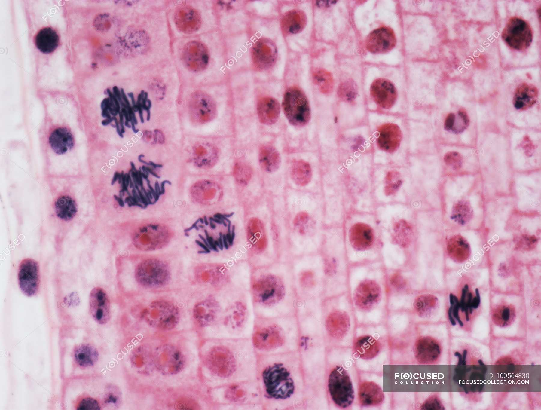
Zwiebelzellen durchlaufen Mitose — Teilung, prophase - Stock Photo |  #160564830
Zwiebelzellen durchlaufen Mitose — Teilung, prophase - Stock Photo | #160564830

Illustrierende Aufgaben zum LehrplanPLUS

Makalah Hasil Pengamatan Sel Bawang Merah | Preparat Mikroskop

Botanische Mikroskopie Zeichnung wo Zeichne ich das Cytoplasma. Allium Cepa?  (Botanik, Zellen, zellkern)
Botanische Mikroskopie Zeichnung wo Zeichne ich das Cytoplasma. Allium Cepa? (Botanik, Zellen, zellkern)
Botanik: Mitose bei der Zwiebelwurzel RESTAURIERT! *

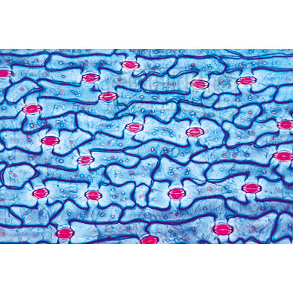
Tulipa, Tulpe, Blattepidermis mit Spaltöffnungen, total / Präparation &  Färbung und Mikroskopierbestecke / Mikroskopie | SCHUCHARDT Lehrmittel
Tulipa, Tulpe, Blattepidermis mit Spaltöffnungen, total / Präparation & Färbung und Mikroskopierbestecke / Mikroskopie | SCHUCHARDT Lehrmittel

Material-is-me: 2015